-
Asked by anon-251613 on 1 May 2020. This question was also asked by anon-253271.
Question: How do you distinguish different cells under the microscope? What do they look like?
- Keywords:
-

Kim Liu answered on 1 May 2020: last edited 1 May 2020 12:04 pm
Different human cells often look different enough under the microscope – whilst they all look vaguely like translucent blobs, different cells are different sizes, different shapes (some have more ‘sharp’ edges, some are more round), some float whilst some are stuck to the dish, and some grow in different patterns (in spheres or branches).
Non human cells like bacteria and fungi can look radically different again, for example bacteria can have tails or grow in rods.
It is also possible to label specific cells with fluorescent markers, so the correct cells light up. This can be done with genetic engineering, or by treating cells with antibodies which detect specific markers on the cell surface.
-

Carolina Coelho answered on 1 May 2020:
they have different sizes and shapes and their insides look different too. Look up histology and you can start to see some of those differences, for example:

-

Delma Childers answered on 1 May 2020:
Yeast look like little rounded eggs under the microscope. Some fungi form these long spindly cells that look a lot like tree branches. Some fungi look like giant swollen spheres. Bacteria also have all sorts of different shapes. Some bacteria form grape-like clusters, some bacteria are long and skinny (bacilli), some are spiral shaped. Human and mouse macrophages are pretty big in comparison to microbes and look round if they’re unhappy or dead or like very flattened triangular-ish shadows.
-

Nina Rzechorzek answered on 1 May 2020:
Brilliant question again! Yes, we can use the shape of the cells under a standard light microscope to help us distinguish between different cell types. However a more powerful way to do this is to use a fluorescent microscope to look for specific ‘cell identity markers’. How do we do that? Well, we know that certain cell types express specific proteins that other cells do not (or at least not in very high amounts). We can use antibodies that bind to those proteins to ‘tag’ the cells we are interested in. We can then use a second antibody that has a coloured fluorescent tag to bind to the first antibody and hey presto – we make different cell types fluoresce with different colours! Here’s a human brain cell called an astrocyte that I took a picture of – fluorescently labelled for GFAP (glial fibrillary acidic protein) which is expressed at high levels in astrocytes:
https://twitter.com/Neurocool/status/519981579507351552/photo/1
And here are some human neurons (the cells in yellow were fluorescently labelled for a neuronal cell marker beta-III tubulin):
https://twitter.com/Neurocool/status/618524578458005504/photo/1 -

Freya Harrison answered on 1 May 2020: last edited 1 May 2020 5:21 pm
Can I add a particularly beautiful example of using fluorescent markers to distinguish different cells? (As Kim and Nina mentioned). This picture shows the different bacteria on a human tongue, coloured with different fluorescent dye molecules. You can see that different bacteria live in different areas of your tongue!
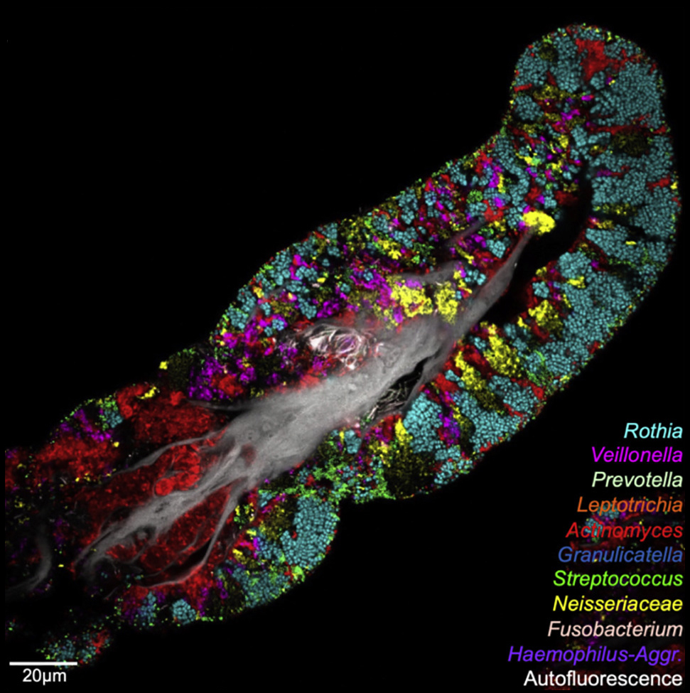
To make this kind of image, scientists make short sequences of DNA that they know will each match a gene in a different species of bacteria. They stick a fluorescent dye molecule on each piece of DNA – a different colour for each sequence. Then they treat the sample they want to look at to make the membranes of bacteria porous. When you add these labelled bits of DNA to your sample, they go into the bacterial cells and the DNA sequences bind to their specific partners – so each bacterial species glows a different colour when you look under the microscope.
-

Luke Bryden answered on 1 May 2020:
As others have said, some cells are different shapes and sizes. However, sometimes it is really hard to distinguish one cell from another. I work with sections of mouse brain tissue, and without some additional steps, determining one cell type from another is really difficult. Distinguishing cell types in the brain is really important because they are all part of different networks and are involved in different functions. They are often intermingled, so we need ‘markers’ to distinguish one cell type from another.
For example, I study a group of nerve cells that produce the chemical messenger dopamine. Dopamine-producing neurons express an enzyme called tyrosine hydroxylase, which is involved in making dopamine. I use a technique called immunohistochemistry to visualise the dopamine-producing nerve cells. Firstly, I add a specially-engineered type of antibody to the brain sections that bind selectively to tyrosine hydroxylase (called the primary antibody). These are similar to the antibodies found in your immune system, but have been engineered to bind to tyrosine hydroxylase rather than to proteins found on, for example, viruses. Next, I add an antibody designed to bind to the primary antibody (these are called secondary antibodies). These secondary antibodies help me to visualise the dopamine-producing nerve cells because they emit a fluorescent signal, which I can detect with a microscope!
––––––––––––––––––––––Ta-da!:

––––––––––––––––––––––
Any neurons glowing – because of the fluorescence – contain tyrosine hydroxylase. And in the part of the brain I am interested in, I know that this means they are dopamine-producing nerve cells!

Comments
Eleanor commented on :
When I learned to use a microscope first, I was mainly able to see pink and purple blobs and wondered how people can distinguish different cell types. But as you learn more, it is possible to distinguish cell type and tissues by the patterns they form. Different colours can be used to highlight parts or types of cells. Research scientists use this to look at whole tissues, individual cells or parts of cells and find out how they work. Clinical scientists (histopathologists) use this to diagnose whether lumps removed from people are cancerous or benign (not a problem) or to identify infections or other disease processes.